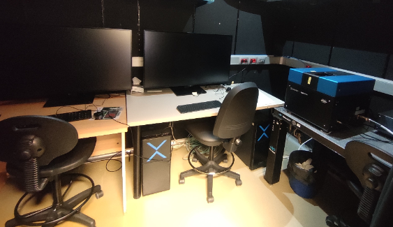
Bruker SPIM LCS
SPIM microscopy is an optical sectioning technique in which the illumination and detection optical paths are perpendicular to each other. The sample is illuminated through a thin light sheet to excite the sample, the detector lens is perpendicular to the illumination path (in this case in a vertical configuration). As a result, optical sections are obtained because only the fluorescent molecules of the focal plane are illuminated. Furthermore, the illumination and detection of each section are done at once, minimizing the light dose applied to the sample and achieving rapid acquisition. The added benefit of this technique is that all the light used to illuminate the sample is used to excite the acquired section, with the double advantage of reducing the damage caused by the excitation light to the fluorochromes and increasing the acquisition speed compared to confocal microscopy (between 100 and 1000 times faster). When used in combination with tissue clarification techniques, SPIM microscopy can detect deep fluorescent signals from large biological samples (greater than 2cm3) with excellent spatial coverage and high temporal resolution.
| Applications | Compact single plane illumination system, designed for rapid 3D capture of clarified samples |
|---|---|
| Software |
|
| Objectives |
|
| Incubator | No |
| Sample Setup | Sample holders cuvette sizes [length x width x height]:
|
| Illumination Source |
|
| Detectors/Cameras | Hamamatsu Orca Flash 4.0 |
| Filters |
|


