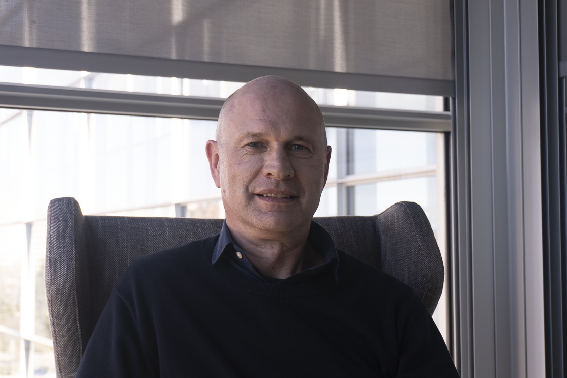
Paul Rilay: "I’m optimistic about the future"
British Heart Foundation
Paul Riley is the British Heart Foundation Personal Chair of Regenerative Medicine at the University of Oxford. He is also Director of the BHF Oxbridge Centre for Regenerative Medicine. He was Professor of Molecular Cardiology at the UCL-Institute of Child Health, London, where he was a principal investigator within the Molecular Medicine Unit at UCL-ICH for 12 years. He obtained his PhD from University College London and completed post-doctoral fellowships in Toronto and Oxford. In 2008, Professor Riley was awarded the Outstanding Achievement Award of the European Society of Cardiology in recognition of the discovery that epicardial cells can regenerate the heart of adult mammals, and in 2014 he was named member of the United Kingdom’s Academy of Medical Sciences. His research interests cover diverse aspects of cardiovascular development, and the mechanisms involved in restoring embryonic potential in the adult heart with disease or lesions in order to promote optimal repair and regeneration.
- Your work focusses on regenerative medicine, particularly in the heart. Could you summarize the current state of your research?
There are currently a combination of approaches in regenerative medicine for the heart. One of them is the transplant of cells or tissue engineering to repair the lesion by introducing new cell types. However, this approach has not met much success because many transplanted cells do not survive or correctly integrate. On the other hand, keeping the patches alive and vascularized is also a challenge. Another focus consists of stimulating endogenous cell types designed to respond to a lesion and promote the growth of the heart muscle from existing muscle that survives the lesion or foster the growth of new blood vessels to support the new muscle. This also includes modulating inflammatory response and fibrosis. These processes can potentially be boosted by activation of cells using molecules or nucleic acid therapeutics such as modified RNA (similar to the COVID vaccine).
- Many organs cannot repair themselves.
If you have a heart attack and lose a significant portion of your vasculature and heart muscle, you risk developing heart failure, which is a debilitating disease that affects 65 million people worldwide. Heart failure has an awful prognosis, significantly reduces quality of life and is, ultimately, fatal. The aim of regenerative medicine is to prevent this by restoring new cells and tissues in the heart and tackling heart failure itself with reversion of the fibrosis or promotion of new blood vessels. This is crucial because heart failure is a common problem worldwide, not just in the western world but increasingly in low and middle-income countries.
- What are the main challenges in this field??
The challenges include achieving the right balance; for instance, when stimulating the growth of heart muscle it is crucial to ensure sufficient vascularization to support the muscle. When reducing fibrosis, there should be heart muscle to replace the scar tissue, which is formed because the heart muscle cannot renew itself. Another great challenge is heart-specific therapeutic delivery. Methods being explored include invasive injections into the heart muscle or blood vessels, but it would be ideal to have a less invasive, oral drug directed at the heart. Safety is also a concern. These drugs should be safe, controllable and specific for the type of heart cells. For instance, large animal studies have shown that stimulating the growth of heart muscle too much can cause arrhythmias and be fatal. So we need therapies that can be turned on and off, which are also safe and effectives.
There are currently a combination of approaches in regenerative medicine for the heart. One of them is the transplant of cells or tissue engineering to repair the lesion by introducing new cell types. However, this approach has not met much success because many transplanted cells do not survive or correctly integrate
- How safe are these therapies?
Studies on animals suggest that they can be safe if they are properly regulated, and the right level of cell regeneration is achieved. However, stem cell transplantation is more difficult to control because many transplanted cells do not survive in the inflammatory, fibrotic environment of a lesioned heart. Patients often need immunosuppressive drugs, which can be toxic. What’s more, we don’t fully understand what drives these cells to achieve clinically beneficial outcomes; they often die shorty after transplant. So using drugs that can be turned on and off, targeted at the types of resident cell, is a more promising approach. The heart is also one of the organs least susceptible to tumours, which makes it a relatively safe target for such therapies.
- Are there animal models that can regenerate their hearts? What can we learn from them?
Yes, there are several models. For instance, adult zebra fish can completely regenerate its heart in approximately 30 days after the loss of a portion or a tissue lesion. Another model is the neonatal mouse, which can completely repair its heart during the day following its birth if there is a lesion. Studying these models helps us understand the mechanisms underlying cardiac regeneration. It has also been seen that human babies regenerate their hearts after an in utero heart attack, showing functional recovery after birth. These models are vital to help understand how to drive the growth of new heart muscle and blood vessels.
Although they are rare, there are also various documented cases of babies that have managed to regenerate their heart after suffering a heart attack. Babies who suffer an in utero heart attack and are then born and treated successfully have shown full regeneration and functional recovery. Although there are few such cases, they show that human babies can regenerate their hearts and studying them can provide valuable information.
- This shows that we can renew our hearts when we are very young, but we lose the capacity. Do we know why?
Thanks to study of the neonatal mouse model, we know that between the first and seventh day after birth, the muscle cells of the heart mature, get larger and have more than one nucleus, which makes them less able to divide and form new muscle. Changes in signalling and maturation of the immune system during this period improve heart function but reduce regenerative capacity. The heart, designed to beat thousands of millions of times during a life, develops to have minimal cell renewal to maintain precise functioning. In early development, there are more opportunities for the formation of new cells. Understanding these signals and key targets can potentially help us restore this capacity in adults with heart lesions.
- Given your experience, do you think we will achieve the target of cardiac regeneration in the next 10-20 years?
Yes. And studies exist on large animal pre-clinical models that suggest it is possible to boost different aspects of heart regeneration. What we need is a collaborative research centre focussed on the proliferation of heart muscle, new blood vessels, the immune system and fibrosis. This centre should work with industrial partners to develop vectors and controllable, targeted nucleic acid therapies. The centre could achieve a new therapeutic treatment in 7-10 years and take it to clinical trials. I’m optimistic about the future.
- Do you have collaborations with the CNIC??
Yes, I have collaborations with Enrique Lara and an EU Marie Curie programme, studying the neonatal mouse model. I am also familiar with the work of Miguel Torres and José Luis de la Pompa, which is very prominent in cardiovascular development and regeneration.


