Nature Communications: Stem cells regulate their own proliferation and their microenvironment
CNIC researchers has identified a new mechanism through which hematopoietic stem cells (HSCs) control both their own proliferation and the characteristics of the niche that houses them. This control is exercised by the protein E-Selectin Ligand-1 (ESL-1). The research team, led by Drs. Andrés Hidalgo and Magdalena Leiva, detected high expression of ESL-1 in HSCs and also found that it controls their production of the cytokine protein TGF-b. This is important because TGF-b has antiproliferative properties and is essential for impeding the loss of HSCs in some diseases, such as some types of anemia. The study was published in Nature Communications.
The team also showed that cells lacking ESL-1 are resistant to a range of cytotoxic and chemotherapeutic agents. These results suggest that ESL-1 is a potential target for therapies aimed at improving bone marrow regeneration after chemotherapy or for expanding the HSC population in preparation for donation.
Hematopoietic stem cells give rise to all blood cells, and as Dr. Hidalgo explained, these cells “self-renew by making copies of themselves, and at the same time generate blood cells throughout life. These cells include the red blood cells that transport oxygen to all the body’s tissues and all types of white blood cells, which are indispensable for the function of the immune system.” A fundamental characteristic of stem cells is quiescence, the ability to remain at rest without dividing. This ensures a continuous supply of stem cells and their maintenance in disease states that acutely increase the demand for new blood cells or in response to agents that damage DNA, including many chemotherapy drugs.
Microenvironment
Most HSCs are found in the bone marrow, in the interior of bones. Dr. Hidalgo describes how HSCs reside in a “microenvironment” or niche that provides “the necessary elements for their optimal maintenance.” Any perturbation of this niche directly endangers the function of these cells, and can lead to diseases such as leukemias and aplasias. Dr. Hidalgo explains that “we still have much to learn about the mechanisms and cellular components of this medullary microenvironment, and acquiring new knowledge in this area is of vital importance because of its important therapeutic implications".
The researchers identified this pathway by analyzing the bone marrow of animals that lack ESL-1. In the absence of ESL-1, the HSCs proliferate less and are therefore of superior quality and more suitable for potential therapeutic applications. “We see that these cells are resistant to processes associated with bone marrow damage, such as cell death triggered by cytotoxic agents,” comments Dr. Leiva. Indeed, one of the most significant findings of the study is the resistance of stem cells lacking ESL-1 to the deleterious effects of cytotoxic and chemotherapy agents. These results indicate that ESL-1 is a possible “therapeutic target for improved regeneration of the bone marrow during chemotherapy.”

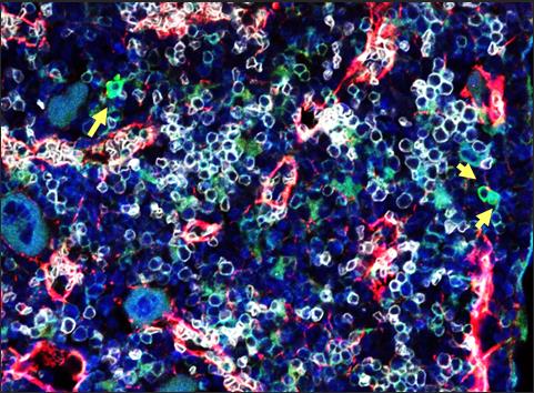
Figura 1. Imagen de microscopia de fluorescencia de la médula ósea de un ratón. Las flechas señalan las células troncales hematopoyéticas.

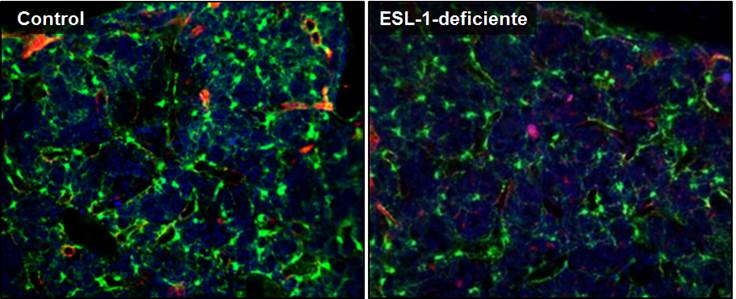
Figura 2. Imágenes de microscopia de fluorescencia de las médulas óseas de ratones reporteros para la citoquina de nicho CXCL12 trasplantados con médula ósea control (izquierda) o ESL-1-deficiente (derecha). El verde (señal GFP) identifica a las células productoras de CXCL12.


