JACC: CNIC and Philips develop ultrafast cardiac magnetic resonance technology that analyzes the heart in less than 1 minute
Ultrafast cardiac magnetic resonance allows precise assessment of heart anatomy and function while reducing healthcare costs and increasing patient comfort
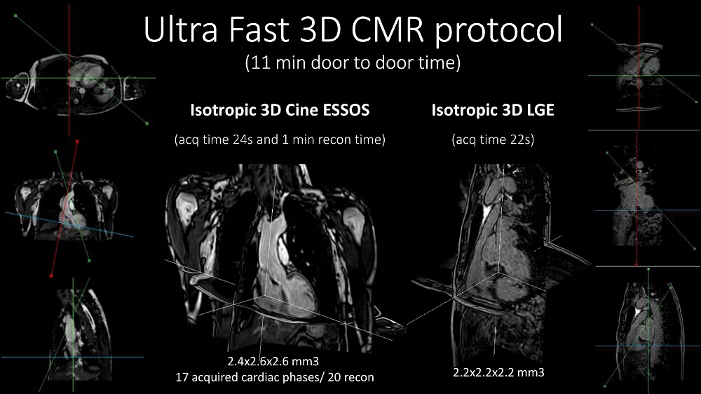
Scientists at the Centro Nacional de Investigaciones Cardiovasculares (CNIC) and Philips have developed a revolutionary technology that can perform cardiac magnetic resonance (CMR) scans in under a minute. ESSOS (Enhanced SENSE by Static Outer volume Subtraction) allows precise assessment of heart anatomy and function, as well as reducing healthcare costs and increasing patient comfort. The new methodology has been tested on more than 100 patients with diverse heart conditions. The results have just been published in JACC: Cardiovascular Imaging, the world-leading journal in the field of cardiac imaging.
CMR provides a noninvasive and radiation-free method for exploring the heart and is the ideal technique for studying heart anatomy, function, and even cell composition. Although most hospitals have magnetic resonance scanners, these are not often used for heart studies because a complete CMR study takes so long. Study first author Dr. Sandra Gómez-Talavera, a CNIC investigator and cardiologist at Fundación Jiménez Díaz University Hospital, explained that “a complete CMR study takes 45-60 minutes, and many patients don’t go through with it because remaining in the scanner for this long is too uncomfortable.”
In addition, hospital magnetic resonance scanners are needed for other studies, limiting their availability for long-duration cardiac studies.
To overcome these obstacles to CMR, CNIC scientists working in partnership with Philips have developed a technique for accelerated CMR acquisition. The technique, explained Dr. Gómez-Talavera, “allows the study of the anatomy and function (motility) of the heart muscle, as well as infarcted and fibrotic tissue. The method can be used to study the whole thoracic cavity in 3D, with algorithms used to focus exclusively on the heart and major vessels (the mobile elements), reducing the scan time.”
The technique, called ESSOS (Enhanced SENSE by Static Outer volume Subtraction), has been tested on more than 100 patients with diverse heart conditions
“We have demonstrated in a large group of patients that CMR with this technology yields the same information as the standard technique, but for less than 10% of the patient scan time,” said Dr. Borja Ibañez, CNIC Clinical Research Director, a cardiologist at Fundación Jiménez Díaz University Hospital, and CIBERCV group leader. Dr. Ibañez is one of the two lead authors on the study.
ESSOS is protected by a patent held jointly by the CNIC and Philips and is the fruit of almost 10 years’ collaboration. The research team believe that this new technology will revolutionize cardiac imaging.
The new study is one of the highlights of the partnership between the CNIC and Philips. Study author Dr. Valentín Fuster, CNIC General Director and Physician un Chief Hospital Mount Sinai Nueva York, emphasized that the agreement signed 10 years ago provided the CNIC with more than just the most up-to-date imaging technology. “Having the most advanced imaging equipment is important, but more important still is having a team of scientific experts in imaging who can get the most of the technology. Philips provide this expertise, and we signed a joint technology development agreement with them that has produced many successes.”
“Our fruitful partnership is based on CNIC scientists’ profound knowledge of what is needed in the field of cardiology and the ability of the Philips team to provide technical solutions to improve that diagnosis and treatment of patients,” said joint study lead author Javier Sánchez-González, the Philips scientist who leads the collaboration and is the brains behind the new technological advance.
ESSOS allows images to be obtained 34 times faster than with the standard technology in current use. “All the information needed to understand heart form and function can be acquired in a little over 20 seconds,” indicated Sánchez-González, adding that “a further 20-second acquisition is all that is needed to detect infarction or fibrosis. This brings the scan to an end, in less than 1 minute.”
A key advantage of the technology is that it can be used with the magnetic resonance scanners already installed in hospitals.
A key advantage of the technology is that it can be used with the magnetic resonance scanners already installed in hospital
The research was funded by the Instituto de Salud Carlos III through a FIS technology development grant, a Spanish Society of Cardiology Translational Research award, the European Research Council (ERC), and the Comunidad de Madrid (Red Madrileña de Nanomedicina en Imagen Molecular).
·


