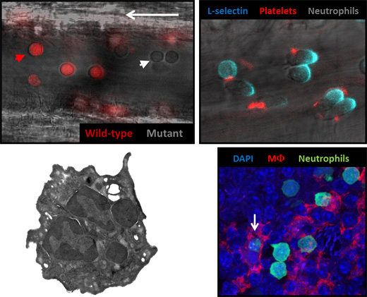
Legend for image panel: (Top left to bottom right) (1) Images from an intravital experiment of the inflamed vasculature of the cremaster muscle, in which we can identify and compare the behavior of leukocytes of different genotype on the basis of endogenous fluorescence, in this case mutant cells are detected by contrast and wild-type cells by expression of DsRed (red). (2) Inflammatory neutrophils (contrast) displaying polarization of the surface receptor L-selectin (blue) capture platelets (red) from the circulation. (3) Electron micrograph of an “aged” neutrophil. (4) Neutrophils physiologically cleared in the spleen of a mouse can be identified by GFP expression (green) in close interaction with red-pulp macrophages (red). Some neutrophils are actively engulfed by tissue macrophages (white arrow).


