News search

E.g., 22/11/2024
E.g., 22/11/2024
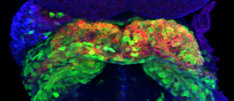
An optical section from a ventral view of a mouse embryo at embryonic day E8.0. The section is overlain with a cast of the three-dimensional shape of the forming heart (red) and the incipient circulatory system (purple).
Research
17 May 2022

The 3D atlas has allowed the scientists to identify the beginning of left–right asymmetry in the heart

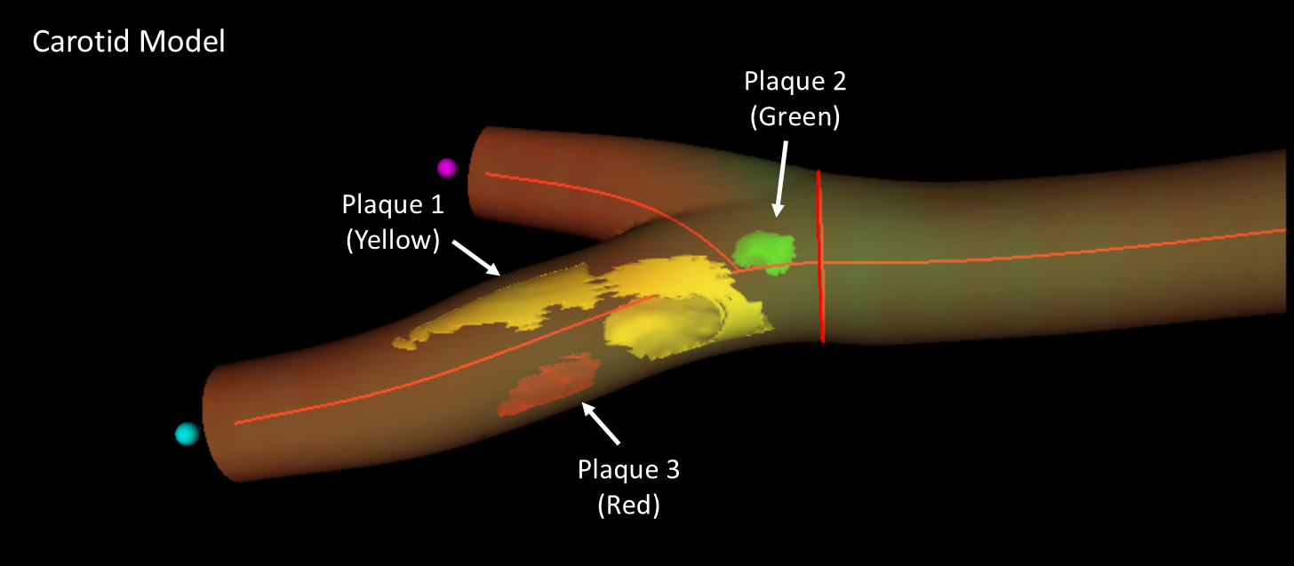
. 3D model of a femoral artery obtained by real 3D imaging, revealing the location, number, and extent of distinct atherosclerotic plaques. In this sample, three plaques can be discerned (arrows), at the femoral bifurcation and distributed along the superficial branch.
Research
16 Mar 2022

Scientists at the CNIC have led the development of a new three dimensional ultrasound method that improves the assessment of cardiovascular risk in healthy individuals


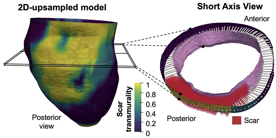
Representative transmurality map from an 3D-upsampled model with a short axis view at high-magnification to illustrate the 3D methodology for transmural-based scar assessment from images obtained with conventional 2D delayed gadolinium-enhanced CMR
Research
28 Sep 2021

This innovative technology may provide an efficient approach in clinical practice after manual or automatic segmentation of myocardial borders in a small number of conventional 2D slices and automatic scar detection


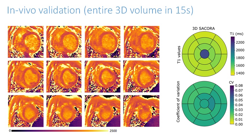
Mapa de T1 obtenido con SACORA y valores promedios
About the CNIC
22 Mar 2021

The development of this 3D T1 mapping technique, called "SACORA", comes as part of the ongoing collaboration between CNIC and Philips

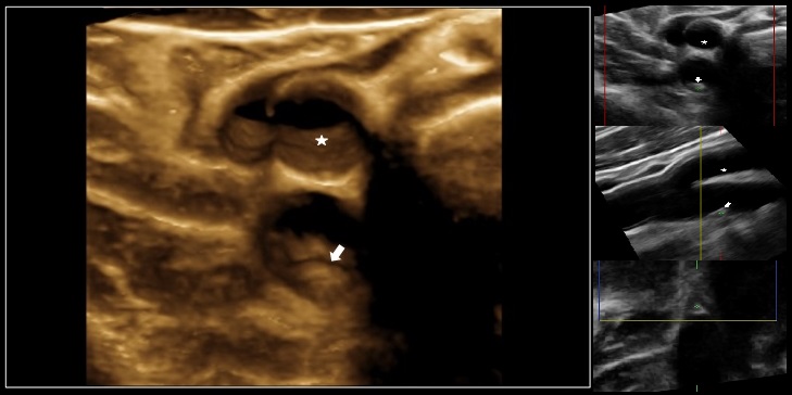
3D ultrasound of the right carotid artery. An atherosclerotic plaque is visible as a small protrusion (arrow) in the vessel wall of the internal branch of the proximal carotid artery (bottom), close to the bifurcation. In contrast, the surface of the disease-free external branch (top) is smooth (asterisk).
Research
24 Jul 2017

CNIC researchers show the value of total atherosclerosis burden for the identification of individuals at risk of cardiovascular disease